

(a)

(b)
C-2
C-1
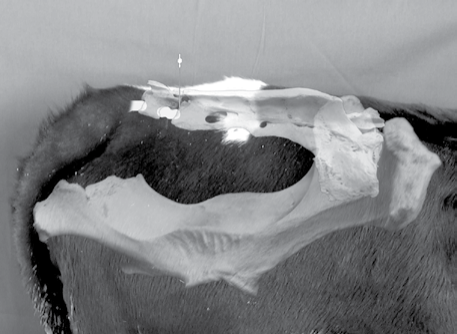
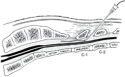
Figure 15.4 (a) Needle placement for epidural. Image courtesy of Dr Douglas Hostetler. (b) Catheter placement for continuous flow epidural. Adapted with permission from Lumb and Jones’ Veterinary Anesthesia .
Sacral paravertebral anesthesia
Sacral paravertebral anesthesia is used to relieve rectal tenesmus associated with rectal prolapse without affecting the sciatic nerve and function of the tail or the animal’s ability to stand. The sacral paravertebral nerve block is used to provide analgesia to the pudendal nerve (pudic nerve), medial hemorrhoidal nerve (pelvic splanchnic nerve), and caudal hemorrhoidal nerve (caudal rectal nerve) by blocking S3, S4, and S5 as they branch off the spinal cord thereby providing analgesia to the anus, vulva, and vagina.1,7 In bulls, S3 supplies motor function to the retractor penis muscles. Physical restraint in a squeeze chute and/or sedation may be beneficial in order to prevent lateral movement of the animal during the procedure. In addition, a caudal epidural may be helpful if the animal is fractious. The skin over the dorsal sacrum should be clipped of hair and surgically prepared for the procedure. The paired S5 foramina are 1–2 cm lateral to the sacral coccygeal joint. The S4 foramina are about 3–4 cm cranial and more lateral to the S5 foramina. The S3 foramina are an additional 3–4 cm cranial to the S4 foramina. A stab incision can be made dorsal to each foramen to aid in the introduction of a 5- to 7-cm 18-gauge needle. The foramina can be palpated rectally with a finger placed in or over the ring which allows for identification of the foramen and ensures correct needle placement. Once the needle has entered the osseous ring, inject 2–3 mL of lidocaine hydrochloride; this should be repeated for each foramen.7 The use of a lidocaine/alcohol mixture has also been described to manage tenesmus following chronic cervicovaginal prolapse or rectal prolapse. A mixture of 1 mL of 2% lidocaine hydrochloride and 2 mL of 95% ethyl alcohol has been used effectively.7
Anesthesia of the penis and prepuce Desensitization of the internal pudendal nerve block
The procedure for bilateral internal pudendal (pudic) nerve block was first described by Larson8 to facilitate relaxation of the bull’s penis without causing locomotor impairment. The internal pudendal nerve block can be used in the
standing bull for penile relaxation and analgesia distal to the sigmoid flexure and examination of the penis. In the standing female, the internal pudendal nerve block can be used to relieve straining caused by chronic vaginal prolapse. This technique may also be used for surgical procedures of the penis, such as repair of prolapses, removal of peri-penile tumors, removal of penile papillomas or warts, and other minor surgeries of the penis and prepuce.
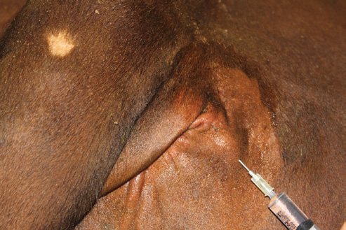
This procedure involves desensitizing the internal pudendal nerve and the anastomotic branch of the middle hemorrhoidal nerve using an ischiorectal approach. The internal pudendal nerve consists of fibers originating from the ventral branches of the third and fourth sacral nerves (S3 and S4) and the pelvic splanchnic nerves. The skin at the ischiorectal fossa on both sides is clipped, disinfected, and desensitized with approximately 2 mL of local anesthetic (Figure 15.5). A 14-gauge 1.25-cm needle is inserted through the desensitized skin at the ischiorectal fossa to serve as a cannula. An 18-gauge 10- to 15-cm spinal needle is then directed through the cannula to the pudendal nerve. The operator’s left hand is placed into the rectum to the level of the wrist and the fingers directed laterally and ventrally to identify the lesser sacrosciatic foramen. The lesser sciatic
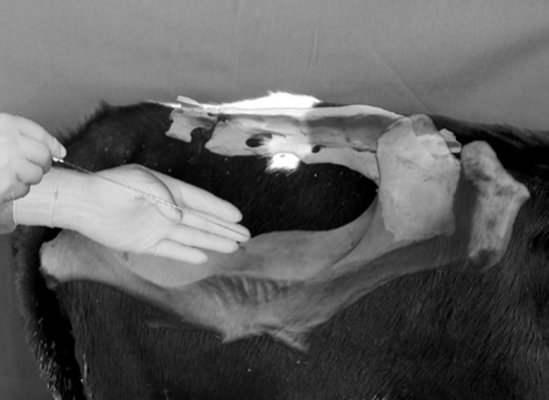
foramen is first identified by rectal palpation as a soft depression in the sacrosciatic ligament. The internal pudendal nerve can be readily identified lying on the ligament immediately cranial and dorsal to the foramen and approximately one finger’s width dorsal to the pudendal artery passing through the foramen. The internal pudendal artery can be readily palpated a finger’s width ventral to the nerve. The spinal needle is held in the operator’s right hand and introduced through the cannula in the ischiorectal fossa. The spinal needle is directed medial to the sacrosciatic ligament and directed cranioventrally (Figure 15.6). The needle is not felt until it has been introduced approximately 5–7 cm and can then be repositioned to the nerve. Once at the pudendal nerve, 20 mL of local anesthetic is deposited at the nerve. The needle is then partially withdrawn and redirected 2–3 cm more caudodorsally where an additional 10 mL of local anesthetic is deposited at the cranial aspect of the foramen to desensitize the muscular branches and the middle hemorrhoidal nerve. The needle is then removed and the sites of deposition are massaged to aid in dispersal of the local anesthetic. This procedure is then repeated on the opposite side of the pelvis. Relaxation of the penis varies and may take as long as 30–40 min for full effect. Effectiveness of the block can be assessed by firmly squeezing the tail of the epididymis of each testicle. The bull’s inability to lift or retract the testicle signifies adequate analgesia. The duration of the internal pudendal nerve block lasts 2–4 hours.7
Desensitization of the dorsal nerve of the penis
The dorsal nerve of the penis may be desensitized at a location just proximal to the surgical site. With the bull restrained, the penis should be manually extended and a towel clamp should then be placed under the dorsal apical ligament. Alternatively, a gauze tourniquet may be placed around the free portion of the penis to aid in penile extension. With the dorsal aspect of the penis thoroughly cleansed, 2–4 mL of 2% lidocaine hydrochloride should be infused subcutaneously across the dorsum of the penis proximal to the lesion.9
Alternatively, the dorsal nerve of the penis may also be desensitized as it passes over the ischial arch for penile
anesthesia and relaxation. The skin associated with the penile body and located 10 cm ventral to the anus and 2.5 cm from midline is infiltrated with 2–4 mL of 2% lidocaine hydrochloride using a small gauge needle (22–25 gauge). A 20-gauge 4-cm needle is then inserted through the desensitized skin and advanced for 5–7 cm to contact the pelvic floor. Aspiration ensures that the needle is not in the dorsal artery of the penis. The needle is then withdrawn approximately 1 cm and the area infiltrated with 20–30 mL of 2% lidocaine hydrochloride. The procedure is then repeated on the opposite side of the penis. Analgesia and paralysis of the penis will occur within 20 min and should last for 1–2 hours.1
Anesthesia for castration
Castration of bulls is a very common surgical procedure in general practice. Historically, castration was often performed with minimal or no anesthesia. However, anesthesia for castration is more commonly practiced because calves benefit from anesthesia with improved feed consumption and rate of gain. Depending on the age and size of the animal, the surgery is usually performed with chemical and/or regional anesthesia (scrotum and testicles). Depending on the size of the calf, the proposed line of incision for removal of the distal aspect of the scrotum should be subcutaneously infiltrated with 5–10 mL of 2% lidocaine hydrochloride. In bulls, an 18-gauge 3.8-cm needle is inserted at an angle (30–45°) into the center of the testicle and 10–15 mL of local anesthetic per 200 kg body weight is injected into the parenchyma of each testicle. The anesthetic quickly enters the lymphatics and desensitizes the sensory fibers in the spermatic cord. For smaller animals or calves, a smaller needle (20 gauge, 2.5 cm) may be used to administer 2–10 mL of 2% lidocaine hydrochloride.1
References
-
Skarda R. Local and regional anesthetic techniques: ruminants and swine. In: Tranquilli WJ, Thurmon JC, Grimm KA (eds) Lumb and Jones’ Veterinary Anesthesia and Analgesia , 4th edn. Oxford: Wiley-Blackwell, 2007, pp. 731–746.
-
Skarda R. Techniques of local analgesia in ruminants and swine. Vet Clin North Am Food Anim Pract 1986;2:621–663.
-
Edwards B. Regional anesthesia techniques in cattle. In Practice 2001;23:142–149.
-
Noordsy J, Ames N. Local and regional anesthesia. In: Noordsy J, Ames N (eds) Food Animal Surgery , 4th edn. Yardley, PA: Veterinary Learning Systems, 2006, pp. 21–42.
-
Noordsy J, Ames N. Epidural anesthesia. In: Noordsy J, Ames N (eds) Food Animal Surgery , 4th edn. Yardley, PA: Veterinary Learning Systems, 2006, pp. 43–55.
-
Navarre C. Numbing: nose to tail. In: Proceedings from the 39th Annual Convention of the American Association of Bovine Practitioners , 2006, pp. 53–55.
-
Hopper R, King H, Walters K, Christiansen D. Management of urogenital surgery and disease in the bull: the scrotum and its contents. Clin Theriogenol 2012;4:332–338.
-
Larson L. The internal pudendal (pudic) nerve block. J Am Vet Med Assoc 1953;123:18–27.
-
Wolfe D, Beckett S, Carson R. Acquired conditions of the penis and prepuce. In: Wolfe DF, Moll HD (eds) Large Animal Urogenital Surgery , 2nd edn. Baltimore: Williams and Wilkins, 1998, pp. 237–272.
Chapter 16
Surgery of the Scrotum and its Contents
Dwight F. Wolfe
Department of Clinical Sciences, College of Veterinary Medicine, Auburn University, Auburn, Alabama, USA
Normal scrotum and testes
The scrotum of the bull is a dependent appendage of the ventral abdominal skin that supports and protects the testicles and helps regulate testicular temperature. The testis is an abdominal organ maintained 2–7 °C cooler than body temperature.1 Testicular thermoregulation is a complex process that includes contraction of the tunica dartos within the scrotal wall to alter scrotal surface area in concert with the cremaster muscles to regulate the distance of the testicles from the body. The scrotum is one of the few places in the bovine where sweat glands are found. Blood temperature is regulated by counter-current heat exchange in the pampiniform plexus just proximal to the testicle between the testicular artery and vein. These mechanisms collectively function to maintain testicular temperature 4–6 °C below core body temperature, which is optimal for normal semen production.1,2
Diagnosis of testicular disease or injury
Bulls with scrotal or testicular disease or injury frequently have scrotal enlargement, which may be unilateral or bilateral and can be caused by a variety of conditions.2,3 It is important to use both visual and palpable information to determine the source of the swelling and make a definitive diagnosis.
The most common cause of scrotal enlargement in the bull is fluid accumulation within the vaginal cavity.3 Fluid accumulation is usually unilateral and may be due to periorchitis, hydrocele, or hematocele. Appreciable fluid accumulation is readily detectable by palpation of the testis; the consistency of the fluid may be thin and easily displaced by palpation, or thicker (e.g., purulent material or clotted blood) which may be confirmed by ultrasound. Additionally, fibrinous or fibrous adhesions may form between the testis or epididymis and parietal vaginal tunic and these may be
detected by palpation as the testis is moved within the vaginal cavity.
Orchitis is usually subclinical but rarely may range to severe and perhaps suppurative.4,5 The dense fibrous tunica albuginea surrounding the testicle limits this organ’s potential for enlargement and therefore there is usually not significant testicular swelling. Although orchitis may affect one or both testes, greater than 25% size difference between the two testes should be considered abnormal. Orchitis in the bovine is usually due to hematogenous spread of bacteria but in rare cases may be due to puncture wounds through the scrotum. Occasionally, traumatic testicular rupture may occur and is diagnosed by an amorphous clot-like consistency by palpation and confirmed by ultrasound.
Epididymitis occurs more commonly than orchitis in bulls and may be diagnosed by thorough examination of the testes and scrotal contents.6–8 The condition is often unilateral in the bull and may present as a swollen painful epididymal tail in the acute phase of the disease. Chronic epididymitis usually results in epididymal tails that are small and firm, and infertility is often caused by their eventual obstruction. Periorchitis – inflammation of the peritesticular tissues with fluid accumulation and adhesions – frequently accompanies epididymitis or orchitis.
Trauma is a common cause of pathological changes in scrotal contents. Trauma to the scrotum may produce hematocele (hemorrhage into the vaginal cavity) or hydrocele (accumulation of inflammatory exudate or transudate in this cavity).3 Trauma severe enough to damage the tubular integrity of the testicle or epididymis may lead to an autoimmune reaction due to the extravasated haploid sperm that produce granulomatous reactions.
Swelling of the wall of the scrotum can usually be differentiated from other causes of scrotal enlargement by palpation of the thickened scrotal skin, presence of pitting edema, with confirmation by ultrasound (Figure 16.1). Generalized swelling of the scrotal wall may be caused by infection with
