
L-4
L-2
L-1
T-13
a
b
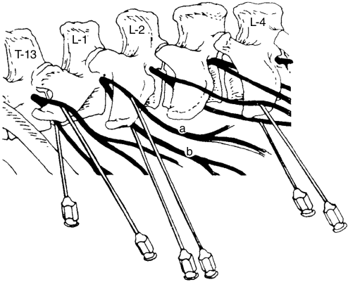
Рисунок 15.3 Дистальная паравертебральная блокада. Воспроизведено с разрешения: Skarda R. Методы местного обезболивания у жвачных животных и свиней. Vet Clin North Am Food Anim Pract 1986;2:635.
и равномерная зона обезболивания и расслабления мышц, снижение внутрибрюшного давления и отсутствие местного анестетика по краям операционной раны. К недостаткам проксимальной паравертебральной блокады относятся искривление позвоночника, которое может затруднить ушивание разреза, сложность определения анатомических ориентиров у животных с ожирением и хорошо развитой мускулатурой, а также необходимость большего опыта и навыков для получения стабильных результатов.2–4
Дистальная паравертебральная блокада
Дистальная паравертебральная блокада обезболивает дорсальные и вентральные ветви спинномозговых нервов T13, L1 и L2 у дистальных концов поперечных отростков L1, L2 и L4 соответственно. Иглу калибра 18 длиной от 3.5 до 5.5–cm вводят вентральнее поперечного отростка и веерообразно распределяют 10 mL местного анестетика. Затем иглу можно полностью извлечь и ввести повторно или изменить её направление, расположив дорсальнее поперечного отростка и направив каудально; там также веерообразно распределяют 10 mL местного анестетика. Эту процедуру повторяют у поперечных отростков второго и четвёртого поясничных позвонков (рисунок 15.3). По сравнению с проксимальной паравертебральной блокадой преимущества дистальной состоят в отсутствии искривления позвоночника, большей простоте выполнения и более стабильных результатах. К её недостаткам по сравнению с проксимальной паравертебральной блокадой относятся необходимость более высоких доз анестетика и различия в эффективности, обусловленные вариациями анатомического хода нервов.2–4
Анестезия промежности
Анестезию промежности обычно проводят при акушерских вмешательствах и для устранения ректальных тенезмов у коров. Она также необходима при многих операциях на мочеполовых органах у коров (например, при вправлении выпадения влагалища или шейки матки) и быков или волов (седалищной уретростомии, ампутации полового члена).
Каудальная эпидуральная анестезия
Каудальная эпидуральная анестезия — простой и недорогой метод обезболивания, широко применяемый у крупного рогатого скота. Высокая каудальная эпидуральная анестезия в крестцово-хвостовом промежутке (S5–Co1) обезболивает крестцовые нервы S2, S3, S4 и S5. Низкая каудальная эпидуральная анестезия в первом межхвостовом промежутке (Co1–Co2) обезболивает крестцовые нервы S3, S4 и S5; при увеличении дозы анестетика его действие может распространяться и на нервы, расположенные краниальнее S2.5 По возможности шерсть следует выстричь, а кожу очистить и продезинфицировать.
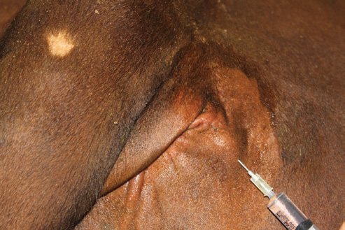
Стоя сбоку от коровы, поднимают и опускают её хвост, чтобы найти углубление между последним крестцовым и первым хвостовым позвонками (первый свободно подвижный промежуток) или между первым и вторым хвостовыми позвонками. Иглу калибра 18 длиной 3.8–cm (без присоединённого шприца) направляют перпендикулярно поверхности кожи. После прокола кожи в канюлю иглы помещают каплю раствора местного анестетика (метод «висячей капли»). Затем иглу медленно продвигают, пока раствор анестетика не втянется в эпидуральное пространство под действием отрицательного давления. После этого к игле присоединяют шприц и медленно вводят раствор анестетика без сопротивления (рисунок 15.4a). Доза местного анестетика составляет 0.5 mL на 45 kg массы тела.
Продлённая каудальная эпидуральная анестезия
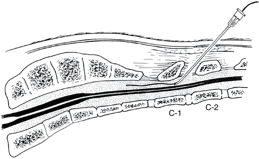
Продлённую каудальную эпидуральную анестезию применяют у крупного рогатого скота с хроническим выпадением прямой кишки или влагалища, если после первоначальной эпидуральной анестезии сохраняются постоянные потуги. Для этого в эпидуральное пространство устанавливают катетер для периодического введения местного анестетика. Спинальную иглу калибра 17 длиной 5–cm (иглу Туохи) с установленным мандреном вводят в эпидуральное пространство между Co1 и Co2, направляя срез краниально. Мандрен извлекают и вводят 2 mL местного анестетика, чтобы проверить нахождение иглы в эпидуральном пространстве. Через иглу вводят катетер и продвигают его краниально на 2–4 cm за кончик иглы. Затем иглу извлекают, оставляя катетер на месте (рисунок 15.4b). На конец катетера устанавливают переходник, а катетер фиксируют к коже на спине. После этого раствор местного анестетика можно вводить по мере необходимости.2
В последнее время для эпидуральной анестезии также применяют агонисты α2-адренорецепторов и опиоиды — отдельно или в сочетании с растворами местных анестетиков. Эпидуральное введение гидрохлорида ксилазина, агониста α2-адренорецепторов, в дозе 0.05 mg/kg, разведённого в 5–12 mL стерильного физиологического раствора, или гидрохлорида ксилазина в дозе 0.3 mg/kg, добавленного к 5 mL 2% гидрохлорида лидокаина, обеспечивает анестезию, сопоставимую с действием лидокаина. Хотя при использовании этих сочетаний продолжительность анестезии увеличивается (4–5 часов), могут возникать и системные эффекты: седация, слюнотечение и атаксия.1 Эпидуральное введение опиоидов, например морфина (0.1 mg/kg), разведённого в 20 mL стерильного физиологического раствора, обеспечивает длительное обезболивание (приблизительно 12 часов), не нарушая двигательной функции. Недостатки применения опиоидов для эпидуральной анестезии состоят в том, что обезболивание выражено слабее, чем при использовании лидокаина, а максимальный эффект эпидурально введённого морфина может наступить лишь через 2–3 часов. Каудальное эпидуральное введение морфина может помочь уменьшить боль в области промежности и потуги.6

(a)

(b)
C-2
C-1
Figure 15.4 (a) Needle placement for epidural. Image courtesy of Dr Douglas Hostetler. (b) Catheter placement for continuous flow epidural. Adapted with permission from Lumb and Jones’ Veterinary Anesthesia .
Sacral paravertebral anesthesia
Sacral paravertebral anesthesia is used to relieve rectal tenesmus associated with rectal prolapse without affecting the sciatic nerve and function of the tail or the animal’s ability to stand. The sacral paravertebral nerve block is used to provide analgesia to the pudendal nerve (pudic nerve), medial hemorrhoidal nerve (pelvic splanchnic nerve), and caudal hemorrhoidal nerve (caudal rectal nerve) by blocking S3, S4, and S5 as they branch off the spinal cord thereby providing analgesia to the anus, vulva, and vagina.1,7 In bulls, S3 supplies motor function to the retractor penis muscles. Physical restraint in a squeeze chute and/or sedation may be beneficial in order to prevent lateral movement of the animal during the procedure. In addition, a caudal epidural may be helpful if the animal is fractious. The skin over the dorsal sacrum should be clipped of hair and surgically prepared for the procedure. The paired S5 foramina are 1–2 cm lateral to the sacral coccygeal joint. The S4 foramina are about 3–4 cm cranial and more lateral to the S5 foramina. The S3 foramina are an additional 3–4 cm cranial to the S4 foramina. A stab incision can be made dorsal to each foramen to aid in the introduction of a 5- to 7-cm 18-gauge needle. The foramina can be palpated rectally with a finger placed in or over the ring which allows for identification of the foramen and ensures correct needle placement. Once the needle has entered the osseous ring, inject 2–3 mL of lidocaine hydrochloride; this should be repeated for each foramen.7 The use of a lidocaine/alcohol mixture has also been described to manage tenesmus following chronic cervicovaginal prolapse or rectal prolapse. A mixture of 1 mL of 2% lidocaine hydrochloride and 2 mL of 95% ethyl alcohol has been used effectively.7
Anesthesia of the penis and prepuce Desensitization of the internal pudendal nerve block
The procedure for bilateral internal pudendal (pudic) nerve block was first described by Larson8 to facilitate relaxation of the bull’s penis without causing locomotor impairment. The internal pudendal nerve block can be used in the
standing bull for penile relaxation and analgesia distal to the sigmoid flexure and examination of the penis. In the standing female, the internal pudendal nerve block can be used to relieve straining caused by chronic vaginal prolapse. This technique may also be used for surgical procedures of the penis, such as repair of prolapses, removal of peri-penile tumors, removal of penile papillomas or warts, and other minor surgeries of the penis and prepuce.
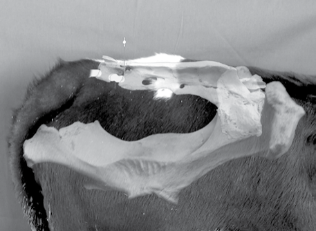
This procedure involves desensitizing the internal pudendal nerve and the anastomotic branch of the middle hemorrhoidal nerve using an ischiorectal approach. The internal pudendal nerve consists of fibers originating from the ventral branches of the third and fourth sacral nerves (S3 and S4) and the pelvic splanchnic nerves. The skin at the ischiorectal fossa on both sides is clipped, disinfected, and desensitized with approximately 2 mL of local anesthetic (Figure 15.5). A 14-gauge 1.25-cm needle is inserted through the desensitized skin at the ischiorectal fossa to serve as a cannula. An 18-gauge 10- to 15-cm spinal needle is then directed through the cannula to the pudendal nerve. The operator’s left hand is placed into the rectum to the level of the wrist and the fingers directed laterally and ventrally to identify the lesser sacrosciatic foramen. The lesser sciatic
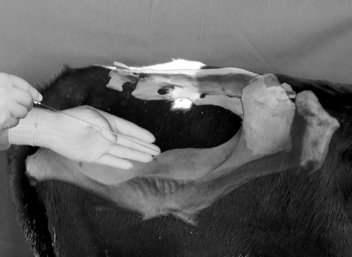
foramen is first identified by rectal palpation as a soft depression in the sacrosciatic ligament. The internal pudendal nerve can be readily identified lying on the ligament immediately cranial and dorsal to the foramen and approximately one finger’s width dorsal to the pudendal artery passing through the foramen. The internal pudendal artery can be readily palpated a finger’s width ventral to the nerve. The spinal needle is held in the operator’s right hand and introduced through the cannula in the ischiorectal fossa. The spinal needle is directed medial to the sacrosciatic ligament and directed cranioventrally (Figure 15.6). The needle is not felt until it has been introduced approximately 5–7 cm and can then be repositioned to the nerve. Once at the pudendal nerve, 20 mL of local anesthetic is deposited at the nerve. The needle is then partially withdrawn and redirected 2–3 cm more caudodorsally where an additional 10 mL of local anesthetic is deposited at the cranial aspect of the foramen to desensitize the muscular branches and the middle hemorrhoidal nerve. The needle is then removed and the sites of deposition are massaged to aid in dispersal of the local anesthetic. This procedure is then repeated on the opposite side of the pelvis. Relaxation of the penis varies and may take as long as 30–40 min for full effect. Effectiveness of the block can be assessed by firmly squeezing the tail of the epididymis of each testicle. The bull’s inability to lift or retract the testicle signifies adequate analgesia. The duration of the internal pudendal nerve block lasts 2–4 hours.7
Desensitization of the dorsal nerve of the penis
The dorsal nerve of the penis may be desensitized at a location just proximal to the surgical site. With the bull restrained, the penis should be manually extended and a towel clamp should then be placed under the dorsal apical ligament. Alternatively, a gauze tourniquet may be placed around the free portion of the penis to aid in penile extension. With the dorsal aspect of the penis thoroughly cleansed, 2–4 mL of 2% lidocaine hydrochloride should be infused subcutaneously across the dorsum of the penis proximal to the lesion.9
Alternatively, the dorsal nerve of the penis may also be desensitized as it passes over the ischial arch for penile
anesthesia and relaxation. The skin associated with the penile body and located 10 cm ventral to the anus and 2.5 cm from midline is infiltrated with 2–4 mL of 2% lidocaine hydrochloride using a small gauge needle (22–25 gauge). A 20-gauge 4-cm needle is then inserted through the desensitized skin and advanced for 5–7 cm to contact the pelvic floor. Aspiration ensures that the needle is not in the dorsal artery of the penis. The needle is then withdrawn approximately 1 cm and the area infiltrated with 20–30 mL of 2% lidocaine hydrochloride. The procedure is then repeated on the opposite side of the penis. Analgesia and paralysis of the penis will occur within 20 min and should last for 1–2 hours.1
Anesthesia for castration
Castration of bulls is a very common surgical procedure in general practice. Historically, castration was often performed with minimal or no anesthesia. However, anesthesia for castration is more commonly practiced because calves benefit from anesthesia with improved feed consumption and rate of gain. Depending on the age and size of the animal, the surgery is usually performed with chemical and/or regional anesthesia (scrotum and testicles). Depending on the size of the calf, the proposed line of incision for removal of the distal aspect of the scrotum should be subcutaneously infiltrated with 5–10 mL of 2% lidocaine hydrochloride. In bulls, an 18-gauge 3.8-cm needle is inserted at an angle (30–45°) into the center of the testicle and 10–15 mL of local anesthetic per 200 kg body weight is injected into the parenchyma of each testicle. The anesthetic quickly enters the lymphatics and desensitizes the sensory fibers in the spermatic cord. For smaller animals or calves, a smaller needle (20 gauge, 2.5 cm) may be used to administer 2–10 mL of 2% lidocaine hydrochloride.1
References
-
Skarda R. Local and regional anesthetic techniques: ruminants and swine. In: Tranquilli WJ, Thurmon JC, Grimm KA (eds) Lumb and Jones’ Veterinary Anesthesia and Analgesia , 4th edn. Oxford: Wiley-Blackwell, 2007, pp. 731–746.
-
Skarda R. Techniques of local analgesia in ruminants and swine. Vet Clin North Am Food Anim Pract 1986;2:621–663.
-
Edwards B. Regional anesthesia techniques in cattle. In Practice 2001;23:142–149.
-
Noordsy J, Ames N. Local and regional anesthesia. In: Noordsy J, Ames N (eds) Food Animal Surgery , 4th edn. Yardley, PA: Veterinary Learning Systems, 2006, pp. 21–42.
-
Noordsy J, Ames N. Epidural anesthesia. In: Noordsy J, Ames N (eds) Food Animal Surgery , 4th edn. Yardley, PA: Veterinary Learning Systems, 2006, pp. 43–55.
-
Navarre C. Numbing: nose to tail. In: Proceedings from the 39th Annual Convention of the American Association of Bovine Practitioners , 2006, pp. 53–55.
-
Hopper R, King H, Walters K, Christiansen D. Management of urogenital surgery and disease in the bull: the scrotum and its contents. Clin Theriogenol 2012;4:332–338.
-
Larson L. The internal pudendal (pudic) nerve block. J Am Vet Med Assoc 1953;123:18–27.
-
Wolfe D, Beckett S, Carson R. Acquired conditions of the penis and prepuce. In: Wolfe DF, Moll HD (eds) Large Animal Urogenital Surgery , 2nd edn. Baltimore: Williams and Wilkins, 1998, pp. 237–272.
